WASSCE Physics · Optical Instruments
The human eye, defects of vision and the camera
By the WASSCE Tutor team
Updated 10 November 2026 · 7 min read · Prepared against the WASSCE syllabus.
Your eye is a living camera: a lens that focuses light onto a light-sensitive screen, with an automatic shutter and focus. Understanding it explains why some people need glasses — and how a camera copies its design.
Learning objectives
By the end of this lesson you should be able to:
- describe the structure of the human eye and the function of each part
- explain accommodation, the near point and the far point
- explain short sight and long sight and how they are corrected
- compare the eye with the camera
The structure of the eye
| Part | Function |
|---|---|
| Cornea | transparent front window; does most of the bending (refraction) of light |
| Iris | coloured ring of muscle that controls the size of the pupil |
| Pupil | the hole through which light enters; smaller in bright light, larger in dim light |
| Eye lens | converging lens of variable thickness; fine-focuses the image |
| Ciliary muscles | change the thickness (focal length) of the lens |
| Suspensory ligaments | hold the lens in place |
| Retina | light-sensitive layer where the image forms; contains rods (dim light) and cones (colour) |
| Fovea (yellow spot) | most sensitive part of the retina, where vision is sharpest |
| Blind spot | where the optic nerve leaves; no light-sensitive cells |
| Optic nerve | carries signals from the retina to the brain |
| Aqueous and vitreous humour | transparent fluids that keep the eye's shape |
The image on the retina is real, inverted and diminished. The brain interprets it as upright.
Accommodation
Accommodation is the ability of the eye to change the focal length of its lens so that objects at different distances are focused clearly on the retina.
- Distant object: ciliary muscles relax, the lens becomes thinner (longer focal length).
- Near object: ciliary muscles contract, the lens becomes thicker (shorter focal length).
- The near point is the closest point at which an object can be seen clearly. For a normal adult eye it is about 25 cm (the least distance of distinct vision).
- The far point is the furthest point that can be seen clearly. For a normal eye it is at infinity.
Defects of vision
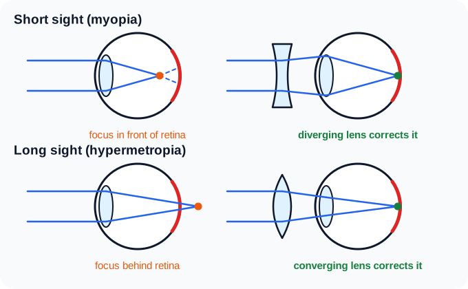
Figure 1. Short sight and long sight, and their correction. WASSCE Tutor diagram.
Short sight (myopia)
- Symptom: near objects are clear; distant objects are blurred. The far point is closer than infinity.
- Cause: the eyeball is too long, or the lens is too strong (converges too much).
- Effect: rays from a distant object focus in front of the retina.
- Correction: a diverging (concave) lens, which spreads the rays slightly before they enter the eye.
Long sight (hypermetropia)
- Symptom: distant objects are clear; near objects are blurred. The near point is further than 25 cm.
- Cause: the eyeball is too short, or the lens is too weak.
- Effect: rays from a near object would focus behind the retina.
- Correction: a converging (convex) lens, which converges the rays a little before they enter the eye.
Other defects
- Presbyopia: with age the lens stiffens and cannot accommodate well, so older people struggle to read; corrected with converging (reading) glasses or bifocals.
- Astigmatism: the cornea is unevenly curved, so lines in some directions are blurred; corrected with cylindrical lenses.
- Cataract: the lens becomes cloudy; treated by surgically replacing the lens.
Free eye-screening camps run by health NGOs in Sierra Leone often find students who cannot read the board from the back of the class — short sight, quickly fixed with diverging spectacles. Cataract surgery restores sight to many older people.
A short-sighted student cannot see clearly beyond 2.0 m. What lens does she need, and what is its power?
She needs a diverging lens that forms a virtual image, at 2.0 m, of very distant objects (). So m:
The eye and the camera compared
| Eye | Camera |
|---|---|
| eye lens and cornea | camera lens (converging) |
| retina | film or digital sensor |
| iris and pupil | diaphragm and aperture |
| eyelid | shutter |
| focusing by changing the thickness of the lens (accommodation) | focusing by changing the distance between lens and film |
| image real, inverted, diminished | image real, inverted, diminished |
| dark choroid layer absorbs stray light | inside of the camera painted black |
Key points
- The cornea and lens focus a real, inverted, diminished image on the retina.
- Accommodation: ciliary muscles change the lens thickness; near point ≈ 25 cm, far point at infinity.
- Short sight: focus in front of the retina; diverging lens. Long sight: focus behind the retina; converging lens.
- The camera focuses by moving the lens; the eye by changing its shape.
WASSCE examination focus
Label a diagram of the eye and give functions of its parts, explain accommodation, explain short and long sight with ray diagrams showing the defect and its correction, and compare the eye and the camera. A calculation of the power of a correcting lens may follow.
Quick check
- Which part of the eye controls how much light enters?
- Which type of lens corrects short sight?
- How does a camera focus objects at different distances?
Answers: 1. The iris (by changing the size of the pupil). 2. A diverging (concave) lens. 3. By moving the lens towards or away from the film or sensor.
Keep going with optical instruments
This is one of 2 lessons in Optical Instruments. Start free with the first lesson of every topic, then unlock them all, plus exam-style practice marked instantly and guided learning tools, from 25 NLe.
- Free account, no card needed
- Everything from 25 NLe
- No automatic renewal
More lessons in Optical Instruments
- Magnifying glass, microscope, telescope and projector🔒